A simple clinical approach to purpura
What is purpura?
Purpura is visible hemorrhage into the skin or mucous membranes due to extravastion of RBCs.
How purpura occurs?
Three possible mechanisms
- Simple hemorrhage.
- Inflammatory hemorrhage (i.e., inflammation directed at the blood vessels)
- Occlusive hemorrhage with minimal inflammation
What are the types of purpura according to morphology?
Six types
- Macular petechiae
- Intermediate macular purpura
- Macular ecchymoses
- Palpable purpura
- Inflammatory retiform purpura
- Non inflammatory retiform purpura
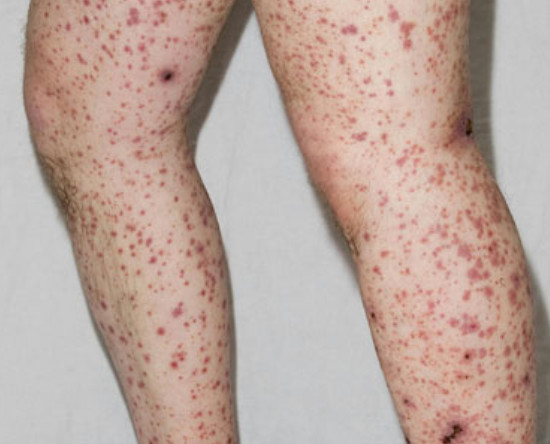
What are petechiae?
Petechiae are purpuric lesions which are
- Small : ≤4 mm in diameter.
- Flat (not palpable).
- Petechiae = simple hemorrhage.

What are the diseases which may present with petechiae?
- Thrombocytopenia (platelets <10000–20000/mm3)
- Abnormal platelet function (hereditary or acquired)
- Pigmented purpuric dermatoses.
- Elevated intravascular pressure spikes.
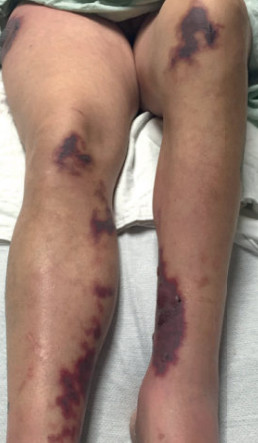
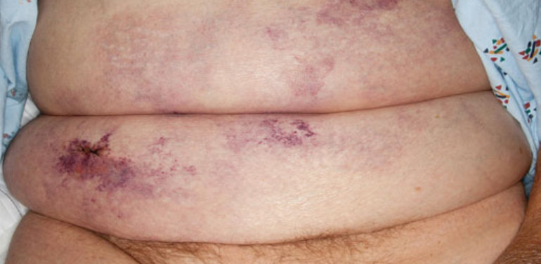
What are Ecchymoses?
Ecchymoses (bruises) are large flat purpuric lesions, 1 cm or more in diameter.
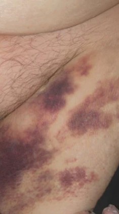
What are the diseases which may present with Ecchymoses?
- Hemophilia
- Anticoagulant use.
- Vitamin K deficiency.
- Hepatic insufficiency.
- Actinic purpura.
- Corticosteroid therapy (topical or systemic),
- Vitamin C deficiency (scurvy).
- Systemic amyloidosis.
- Ehlers- Danlos syndrome.
How to clinically assess the presence of inflammation associated with purpura?
Purpura may be associated with inflammtion By diascopy test or applying pressure on the lesion :
- The blanchable component of the color represents erythema which is a marker for inflammation.
- The non blanchable component of the color represents purpura which is a marker for hemorrhage.
What is Palpable purpura?
Palpable purpura is inflammatory purpura with prominent early erythema partially blanches with diascopy indicating the presence of both inflammation and hemorrhage.
What are the diseases which may present with Palpable purpura?
- Cutaneous small-vessel vasculitis (e.g., leukocytoclastic vasculitis)
- ANCA-associated vasculitides
- Small- and medium-sized vessels` vasculitis.
- IgA vasculitis
- Erythema multiforme.
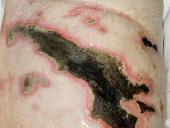
What is retiform purpura?
Retiform means netlike, reticular or branching morphology.
Retiform purpura occurs as a result of complete vascular occlusion and vascular damage involving blood vessels in the skin.
What are the possible causes of inflammatory retiform purpura?
- IgA cutaneous small-vessel vasculitis
- vasculitis affecting both small- and medium-sized vessels (such as ANCA-associated and connective tissue disease-associated vasculitides)
- Chilblains (perniosis).
What are the possible causes of non inflammatory retiform purpura?
- Calciphylaxis.
- Heparin necrosis.
- warfarin necrosis.
- Antiphospholipid antibody syndrome.
- Cryoglobulinemia (monoclonal type only).
- Hypercoagulable disorders (e.g., protein C or S deficiency).
- Disseminated intravascular coagulation.
- Cocaine use (levamisole-tainted).
- Oxalosis.
- Cholesterol emboli
- Livedoid vasculopathy.
- Vessel-invasive infection (e.g., Aspergillus, ecthyma gangrenosum).
- Malignant atrophic papulosis (Degos disease).
- Nonbacterial thrombotic endocarditis.
Approach to purpura
- Is the lesion is purpuric?
- Yes.
- Palpable or not ?
- Palpable : examine early lesions
- Non palpable : classify according to size into
- Macular petechiae: ≤4 mm.
- Intermediate macular purpura: 5-9 mm.
- Macular ecchymoses: ≥1 cm.
- Palpable lesions : according to early lesions examination
- Classic palpable purpura : Early lesions are round, port-wine colored, and have prominent erythema (partially blanch with diascopy).
- Noninflammatory retiform purpura: Early lesions lack erythema and demonstrate a retiform, branched (retiform) appearance, (usually due to conditions associated with microvascular occlusion).
- Inflammatory retiform purpura : Early lesions have prominent erythema and demonstrate a retiform, branched appearance.
- If not sure from the cause of purpura
- Biopsy may be taken from early lesion(24-48 hrs) and direct immunofluorescence study to exclude IgA vasculitis.
- Laboratory investigations may be done based on the morphology of the lesions.
#A simple clinical approach to purpura